The heart and its blood vessels are normally surrounded with a sac-like structure that provides important functions as protection and prevents the heart from over-expanding when there is blood volume excess. The sac is called the pericardium sac, in between the cardiac layers is consists of a fluid called pericardial fluid. This fluid reduces the friction of the pericardium by lubricating the surface of the epicardial. An excessive pericardial fluid is known as pericardial effusion.
A condition in which there is an accumulation of excess fluid around the heart is called pericardial effusion. It may develop as a result of inflammation, trauma, and increase capillary filtration pressure. As the pericardial fluid exceeds the pericardium’s level, it gives pressure to the heart and affects its normal function. If this left untreated, pericardial effusion may lead to a complication called cardiac tamponade – (a life-threatening condition), slow or rapid compression of the heart due to the accumulation of fluid in the pericardial sac. Disturbed equilibrium between the production and re-absorption and structural abnormality is the other cause that allows fluid to enter the pericardial cavity. The normal levels/volume of pericardial fluid are from 15 – 50 mL.
Types of Pericardial Effusion
- Transudative – this due to nephritic syndrome, myxedema, and congestive heart failure.
- Exudative – this due to tuberculosis and empyema – is a collection of pus within a naturally existing anatomical cavity, such as the lung pleura & pericardial sac.
- Hemorrhagic – This due to injury, rupture of aneurysm and malignant effusion.
- Malignant – This is a result of an accumulation of fluid caused by metastasis.
Clinical Manifestations
Small or even large effusion that develops slowly may cause few or no symptoms because pericardium is able to stretch to avoid compression. However, sudden accumulation of fluid of 200 mL may increase the intracardial pressure affecting the heart function. Below are the symptoms of pericardial effusion:
- Dyspnea – a symptom of breathlessness.
- Orthopnea – shortness of breath that occurs when lying flat cough
- Chest pain specifically on the left side of the chest behind the breastbone or sternum. Chest pain usually occurs when breathing.
- Fever due to inflammation.
- Tachycardia or rapid heart rate.
- Fainting and dizziness.
- Ewart’s sign – Dullness to percussion
Causes of Pericardial Effusion
- Inflammation is said to the primary cause of the pericardial effusion. Inflammation in the pericardium or known as pericarditis produces extra fluid resulting in pericardial effusion. Pericarditis may be caused by a bacterial and viral infection. Viruses include coxsackieviruses, HIV, cytomegaloviruses, and echoviruses.
- The disease of connective tissue and trauma due to radiation treatment for cancer may also be caused by pericarditis.
- Virus – HIV; Bacterial – Tuberculosis
- Pericarditis due to unknown causes.
- Sever Cancer that is in the metastatic phase and HIV/AIDS
- Trauma or injury to the pericardium.
- Dressler’s syndrome – a secondary form of pericarditis that results from any type of injury to the heart or pericardium.
- Myocardial infarction (MI) or heart disease
- Autoimmune diseases such as arthritis and systemic lupus erythematosus.
- Radiation therapy and chemotherapy treatment for cancer.
- Severe kidney failure.
- Hypothyroidism
- Medications including Minoxidil, hydralazine, isoniazid, and phenytoin.
Complication
In some cases, pericardial effusion produces no symptoms even a large effusion because pericardium can able to adjust or stretch to avoid compressing the heart. Cardiac Tamponade is the complication if pericardial effusion which describes as a life-threatening, slow or rapid compression of the heart due to the accumulation of pus, fluid, or blood in the pericardium.
Pericardial Effusion Test and Diagnosis
- Blood Test – Complete Blood Count
- Cancer biomarkers are used to predict the natural course of the tumor, suggesting that the patient’s outcome is likely to be good or bad (prognosis).
- Tuberculin test – PPD tuberculin (purified protein derivative tuberculin) is a sterile solution of a purified protein fraction precipitated from a filtrate of tubercle bacillus culture grown on a special medium is use during tuberculin test. A positive result in this test indicates that you have a previous or current infection with Mycobacterium tuberculosis.
- Serum electrolytes.
- Cardiac enzymes
- Thyroid function
- Test for rickettsial antibodies
Chest X-ray
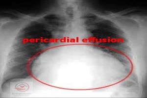
Chest X-ray is a form of electromagnetic radiation that is commonly used as an early part of the diagnostic procedure to view the affected part. Water-bottle like appearance is showed after the test if effusion occurred. However, it is not a reliable test for confirming the diagnosis.
MRI –
MRI or Magnetic resonance imaging device is more sensitive compared to other machines and has no radiation. MRI can accurately detect pericardial effusion even as small as 30 mL. It uses a computer to produce images of the body structures with the help of magnetism, radio waves.
Echocardiography –
Known as echocardiography or cardiac ultrasound, it works on a two-dimensional technique to produce a heart image. And the latest ultrasound system works on 3D real-time imaging. This procedure can provide the accurate size and location of the effusion, and also help health care providers what approach to be adopted in treatment.
CT Scan –
Computed tomography (CT) scan is an advanced machine used to detect pericardial effusion and it is more accurate compared to echocardiography because it can provide 100 times a visible image of the entire body. CT scan uses the same dosage of radiation as that of an ordinary X-ray machine.
Pericardioscopy –
It is allowed to assess and visualization of all surfaces of the pericardium. and can be made easy to perform a selective biopsy, including but not limited to a subxiphoid window. This is a safe procedure that may allow a distinction between benign, tuberculous and malignant origins of pericardial effusion.
Electrocardiogram (ECG) –
The ECG is a noninvasive test that is used to reflect the state of the heart by measuring its electrical activity. ECG changes are prominent in pericardial effusion resulting from pericarditis and Dressler’s syndrome. This used electrical sensing devices or leads to reading the cardiac status.
Pericardiocentesis –
Pericardiocentesis is a procedure where the fluid is aspirated from the pericardium using a needle. This can be done either for diagnostic or for therapeutic reasons. For diagnostic purposes, pericardial fluid is obtained to determine the cause of the effusion. The aspirated fluid may contain blood if it is caused by trauma and myocardial infarction. The rupture of the thoracic duct produces milky fluids. If pericardial effusion caused by bacteria, it produces pus or purulent fluids. Tuberculosis and neoplastic disease produce serosanguineous – composed of serum and blood.
Pericardial Effusion Treatment and Management
Treatment of pericardial effusion depends on how worst the condition is and how much the amount of fluid has accumulated in the pericardium causing a complication called cardiac tamponade. Cardiac tamponade is the worst complication of pericardial effusion where the normal function of the heart is affected due to pressure produced by fluid accumulation.
The symptoms include cyanosis, blue tinge to the lips and skin, shock and change in mental status. This requires urgent drainage of the fluid which accomplished with a procedure called pericardiocentesis – insertion of a needle into the pericardial space and fluid is withdrawn using a large syringe. Treating the underlying cause like pericarditis often corrects the problem.
Pharmacological Treatment
- Anti-inflammatory Drugs – This is usually prescribed to treat the inflammation that contributes to pericardial effusion.
- NSAIDs or Nonsteroidal Anti-inflammatory drugs – such as ibuprofen and indomethacin.
- Aspirin – also known as acetylsalicylic acid. A salicylate type of drug that used as an analgesic to relieve minor pains, reduce fever, and offer anti-inflammatory action.
- Colchicine Drugs.
- Antibiotic medication is used if the underlying cause is due to infection.
- Heart failure medications and Diuretics drugs may be prescribed to treat pericardial effusion.
- Corticosteroid medications are usually prescribed if you don’t respond with the above treatments.
Surgical Treatment
An invasive procedure is the other option to treat pericardial effusion if anti-inflammatory doesn’t correct the problem. Below are the following procedures recommended by the doctor to drain and prevent recurring from fluid accumulation.
Open-Heart Surgery – This invasive produce allows fluids to drain and at the same time may repair any related heart damage due to recent heart surgery.
Intrapericardial Sclerosis – In this procedure, your surgeon will inject a solution between the two layers to seals the layers together.
Pericardiectomy – Is the surgical removal of part or most of the pericardium.
Pericardiocentesis – Is produce where the pericardial fluid is aspirated or drained from the pericardium. In most cases, to prevent reaccumulation of fluid the catheter is left in place to drain the pericardial space.
For More Read About Pericardial Effusion Click Here